-
MERISTEMATIC TISSUE
MERISTEMATIC TISSUE
Activity 6.1:
* Take two glass jars and fill them with water.
* Now, take two onion bulbs and place one on each jar, as shown in Fig. 6.1.
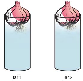
Figure 6.1: Growth of roots in onion bulbs
* Observe the growth of roots in both the bulbs for a few days.
* Measure the length of roots on day 1, 2 and 3.
* On day 4, cut the root tips of the onion bulb in jar 2 by about 1 cm.
After this, observe the growth of roots in both the jars and measure their lengths each day for five more days and record the observations in tables, like the table below:

The growth of plants occurs only in certain specific regions. This is because the dividing tissue, also known as meristematic tissue, is located only at these points. Depending on the region where they are present, meristematic tissues are classified as apical, lateral and intercalary (Fig. 6.2). New cells produced by meristem are initially like those of meristem itself, but as they grow and mature, their characteristics slowly change and they become differentiated as components of other tissues.
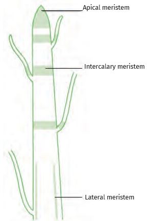
Figure 6.2: Location of meristematic tissue in the plant body
Apical meristem is present at the growing tips of stems and roots and increases the length of the stem and the root. The girth of the stem or root increases due to lateral meristem (cambium). Intercalary meristem seen in some plants is located near the node.
Cells of meristematic tissue are very active, they have dense cytoplasm, thin cellulose walls, and prominent nuclei. They lack vacuoles. Can we think why they would lack vacuoles? (You might want to refer to the functions of vacuoles in the chapter on cells.)
Questions
1. Which of the two onions has longer roots? Why?
2. Do the roots continue growing even after we have removed their tips?
3. Why would the tips stop growing in jar 2 after we cut them?
4. Name types of simple tissues.
5. Where is apical meristem found?
Source: This topic is taken from NCERT TEXTBOOK
-
PERMANENT TISSUE – SIMPLE PERMANENT TISSUE
PERMANENT TISSUE – SIMPLE PERMANENT TISSUE
What happens to the cells formed by meristematic tissue? They take up a specific role and lose the ability to divide. As a result, they form a permanent tissue. This process of taking up a permanent shape, size, and function is called differentiation. Differentiation leads to the development of various types of permanent tissues.
Activity 6.2: (Observing simple permanent tissue)

Figure 6.3: Section of a stem
* Take a plant stem and with the help of your teacher cut into very thin slices or sections.
* Now, stain the slices with safranin. Place one neatly cut section on a slide, and put a drop of glycerine.
* Cover with a cover-slip and observe under a microscope.
* Observe the various types of cells and their arrangement.
* Compare it with Fig. 6.3.
* Now, answer the following on the basis of your observation:
1. Are all cells similar in structure?
2. How many types of cells can be seen?
3. Can we think of reasons why there would be so many types of cells?
* We can also try to cut sections of plant roots. We can even try cutting sections of root and stem of different plants
A few layers of cells beneath the epidermis are generally simple permanent tissue. Parenchyma is the most common simple permanent tissue. It consists of relatively unspecialised cells with thin cell walls. They are living cells. They are usually loosely arranged, thus large spaces between cells (intercellular spaces) are found in this tissue (Fig. 6.4 a). This tissue generally stores food.
In some situations, it contains chlorophyll and performs photosynthesis, and then it is called chlorenchyma. In aquatic plants, large air cavities are present in parenchyma to help them float. Such a parenchyma type is called aerenchyma.
The flexibility in plants is due to another permanent tissue, collenchyma. It allows bending of various parts of the plant-like tendrils and stems of climbers without breaking. It also provides mechanical support. We can find this tissue in leaf stalks below the epidermis. The cells of this tissue are living, elongated and irregularly thickened at the corners. There is very little intercellular space (Fig. 6.4 b).
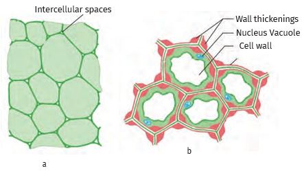
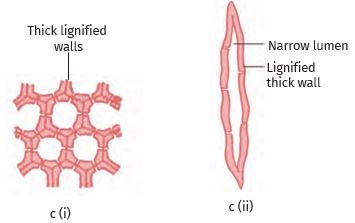
Figure 6.4: Various types of simple tissues: (a) Parenchyma (b) Collenchyma (c) Sclerenchyma (i) transverse section, (ii) longitudinal section
Yet another type of permanent tissue is sclerenchyma. It is the tissue which makes the plant hard and stiff. We have seen the husk of a coconut. It is made of sclerenchymatous tissue. The cells of this tissue are dead. They are long and narrow as the walls are thickened due to lignin. Often these walls are so thick that there is no internal space inside the cell (Fig. 6.4 c). This tissue is present in stems, around vascular bundles, in the veins of leaves and in the hard covering of seeds and nuts. It provides strength to the plant parts.
Activity 6.3: (Observing Guard and Epidermal cells)
* Take a freshly plucked leaf of Rhoeo. Stretch and break it by applying pressure.
* While breaking it, keep it stretched gently so that some peel or skin projects out from the cut.
* Remove this peel and put it in a petri dish filled with water.
* Add a few drops of safranin.
* Wait for a couple of minutes and then transfer it onto a slide. Gently place a coverslip over it.
* Observe under a microscope.
What you observe is the outermost layer of cells, called the epidermis. The epidermis is usually made of a single layer of cells. In some plants living in very dry habitats, the epidermis may be thicker since protection against water loss is critical. The entire surface of a plant has an outer covering epidermis. It protects all the parts of the plant. Epidermal cells on the aerial parts of the plant often secrete a waxy, water-resistant layer on their outer surface. This aids in protection against loss of water, mechanical injury and invasion by parasitic fungi. Since it has a protective role to play, cells of epidermal tissue form a continuous layer without intercellular spaces. Most epidermal cells are relatively flat. Often their outer and side walls are thicker than the inner wall.
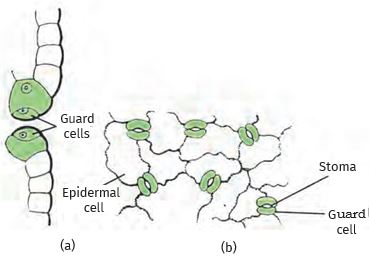
Figure 6.5: Guard cells and epidermal cells: (a) lateral view, (b) surface view
We can observe small pores here and there in the epidermis of the leaf. These pores are called stomata (Fig. 6.5). Stomata are enclosed by two kidney-shaped cells called guard cells. They are necessary for exchanging gases with the atmosphere. Transpiration (loss of water in the form of water vapour) also takes place through stomata.
epidermal cells of the roots, whose function is water absorption, commonly bear long hair-like parts that greatly increase the total absorptive surface area.
In some plants like desert plants, the epidermis has a thick waxy coating of cutin (chemical substance with waterproof quality) on its outer surface. Can we think of a reason for this?
Is the outer layer of a branch of a tree different from the outer layer of a young stem? As plants grow older, the outer protective tissue undergoes certain changes. A strip of secondary meristem located in the cortex forms layers of cells that constitute the cork. Cells of cork are dead and compactly arranged without intercellular spaces (Fig. 6.6). They also have a substance called suberin in their walls that makes them impervious to gases and water.
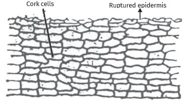
Figure 6.6: Protective tissue
Source: This topic is taken from NCERT TEXTBOOK
-
PERMANENT TISSUE – COMPLEX PERMANENT TISSUE
PERMANENT TISSUE – COMPLEX PERMANENT TISSUE
The different types of tissues we have discussed until now are all made of one type of cell, which looks like each other. Such tissues are called simple permanent tissue. Yet another type of permanent tissue is complex tissue. Complex tissues are made of more than one type of cell. All these cells coordinate to perform a common function. Xylem and phloem are examples of such complex tissues. They are both conducting tissues and constitute a vascular bundle. Vascular tissue is a distinctive feature of complex plants, one that has made possible their survival in the terrestrial environment. In Fig. 6.3 showing a section of stem, can you see different types of cells in the vascular bundle?
Xylem consists of tracheids, vessels, xylem parenchyma (Fig. 6.7 a,b,c) and xylem fibres. Tracheids and vessels have thick walls, and many are dead cells when mature. Tracheids and vessels are tubular structures. This allows them to transport water and minerals vertically. The parenchyma stores food. Xylem fibres are mainly supportive in function.
Phloem is made up of five types of cells: sieve cells, sieve tubes, companion cells, phloem fibres and the phloem parenchyma [Fig. 6.7 (d)]. Sieve tubes are tubular cells with perforated walls. Phloem transports food from leaves to other parts of the plant. Except for phloem fibres, other phloem cells are living cells.
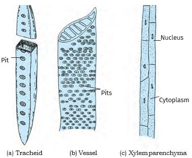
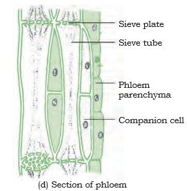
Figure 6.7: Types of complex tissue
Questions
1. Which tissue makes up the husk of coconut?
2. What are the constituents of phloem?
Source: This topic is taken from NCERT TEXTBOOK