-
WHAT ARE CELL ORGANELLES?
WHAT ARE CELL ORGANELLES?
Every cell has a membrane around it to keep its own contents separate from the external environment. Large and complex cells, including cells from multicellular organisms, need a lot of chemical activities to support their complicated structure and function. To keep these activities of different kinds separate from each other, these cells use membrane-bound little structures (or ‘organelles’) within themselves. This is one of the features of the eukaryotic cells that distinguish them from prokaryotic cells. Some of these organelles are visible only with an electron microscope.
We have talked about the nucleus in a previous section. Some important examples of cell organelles which we will discuss now are:
1. endoplasmic reticulum,
2. Golgi apparatus,
3. lysosomes,
4. mitochondria
5. plastids and
6. vacuoles
They are important because they carry out some very crucial functions in cells.
Questions
1. Can you name the two organelles we have studied that contain their own genetic material?
2. If the organisation of a cell is destroyed due to some physical or chemical influence, what will happen?
3. Why are lysosomes known as suicide bags?
4. Where are proteins synthesised inside the cell?
Source: This topic is taken from NCERT TEXTBOOK
-
ENDOPLASMIC RETICULUM
ENDOPLASMIC RETICULUM
The endoplasmic reticulum (ER) is a large network of membrane-bound tubes and sheets. It looks like long tubules or round or oblong bags (vesicles). The ER membrane is similar in structure to the plasma membrane. There are two types of ER– rough endoplasmic reticulum (RER) and smooth endoplasmic reticulum (SER). RER looks rough under a microscope because it has particles called ribosomes attached to its surface. The ribosomes, which are present in all active cells, are the sites of protein manufacture. The manufactured proteins are then sent to various places in the cell depending on need, using the ER. The SER helps in the manufacture of fat molecules, or lipids, important for cell function. Some of these proteins and lipids help in building the cell membrane. This process is known as membrane biogenesis. Some other proteins and lipids function as enzymes and hormones. Although the ER varies greatly in appearance in different cells, it always forms a network system.
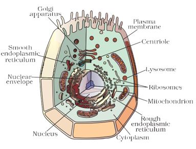
Figure 5.5: Animal cell
Thus, one function of the ER is to serve as channels for the transport of materials (especially proteins) between various regions of the cytoplasm or between the cytoplasm and the nucleus. The ER also functions as a cytoplasmic framework providing a surface for some of the biochemical activities of the cell. In the liver cells of the group of animals called vertebrates, SER plays a crucial role in detoxifying many poisons and drugs.
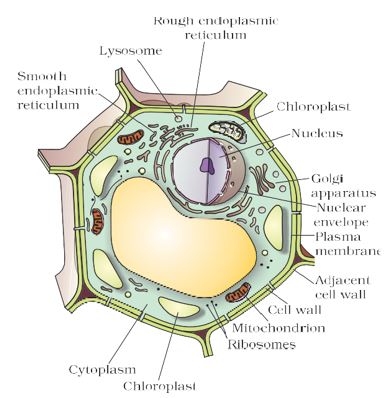
Figure 5.6: Plant cell
Source: This topic is taken from NCERT TEXTBOOK
-
GOLGI APPARATUS
GOLGI APPARATUS
The Golgi apparatus, first described by Camillo Golgi, consists of a system of membrane-bound vesicles (flattened sacs) arranged approximately parallel to each other in stacks called cisterns. These membranes often have connections with the membranes of ER and therefore constitute another portion of a complex cellular membrane system.
The material synthesised near the ER is packaged and dispatched to various targets inside and outside the cell through the Golgi apparatus. Its functions include the storage, modification, and packaging of products in vesicles. In some cases, complex sugars may be made from simple sugars in the Golgi apparatus. The Golgi apparatus is also involved in the formation of lysosomes.
More to know

Camillo Golgi was born at Corteno near Brescia in 1843. He studied medicine at the University of Pavia. After graduating in 1865, he continued to work in Pavia at the Hospital of St. Matteo. At that time most of his investigations were concerned with the nervous system, In 1872 he accepted the post of Chief Medical Officer at the Hospital for the Chronically Sick at Abbiategrasso. He first started his investigations into the nervous system in a little kitchen of this hospital, which he had converted into a laboratory. However, the work of greatest importance, which Golgi carried out was a revolutionary method of staining individual nerve and cell structures. This method is referred to as the ‘black reaction’. This method uses a weak solution of silver nitrate and is particularly valuable in tracing the processes and most delicate ramifications of cells. All through his life, he continued to work on these lines, modifying and improving this technique. Golgi received the highest honours and awards in recognition of his work. He shared the Nobel prize in 1906 with Santiago Ramony Cajal for their work on the structure of the nervous system.
Source: This topic is taken from NCERT TEXTBOOK
-
LYSOSOMES
LYSOSOMES
Structurally, lysosomes are membrane-bound sacs filled with digestive enzymes. These enzymes are made by RER. L ysosomes are a kind of waste disposal system of the cell. These help to keep the cell clean by digesting any foreign material as well as worn-out cell organelles. Foreign materials entering the cell, such as bacteria or food, as well as old organelles end up in the lysosomes, which break complex substances into simpler substances. Lysosomes are able to do this because they contain powerful digestive enzymes capable of breaking down all organic material. During the disturbance in cellular metabolism, for example, when the cell gets damaged, lysosomes may burst and the enzymes digest their own cell. Therefore, lysosomes are also known as the ‘suicide bags’ of a cell.
Source: This topic is taken from NCERT TEXTBOOK
-
MITOCHONDRIA
MITOCHONDRIA
Mitochondria are known as the powerhouses of the cell. Mitochondria have two membrane coverings. The outer membrane is porous while the inner membrane is deeply folded. These folds increase surface area for ATP- generating chemical reactions. The energy required for various chemical activities needed for life is released by mitochondria in the form of ATP (Adenosine triphopshate) molecules. ATP is known as the energy currency of the cell. The body uses energy stored in ATP for making new chemical compounds and for mechanical work.
Mitochondria are strange organelles in the sense that they have their own DNA and ribosomes. Therefore, mitochondria are able to make some of their own proteins.
Source: This topic is taken from NCERT TEXTBOOK
-
PLASTIDS AND VACUOLES
PLASTIDS AND VACUOLES
Plastids
Plastids are present only in plant cells. There are two types of plastids – chromoplasts (coloured plastids) and leucoplasts (white or colourless plastids).
Chromoplasts containing the pigment chlorophyll are known as chloroplasts. Chloroplasts are important for photosynthesis in plants. Chloroplasts also contain various yellow or orange pigments in addition to chlorophyll. Leucoplasts are primarily organelles in which materials such as starch, oils and protein granules are stored.
The internal organisation of the Chloroplast consists of numerous membrane layers embedded in a material called the stroma. These are similar to mitochondria in external structure. Like the mitochondria, plastids also have their own DNA and ribosomes.
Vacuoles
Vacuoles are storage sacs for solid or liquid contents. Vacuoles are small-sized in animal cells while plant cells have very large vacuoles. The central vacuole of some plant cells may occupy 50-90% of the cell volume.
In plant cells, vacuoles are full of cell sap and provide turgidity and rigidity to the cell. Many substances of importance in the life of the plant cell are stored in vacuoles. These include amino acids, sugars, various organic acids and some proteins. In single-celled organisms like Amoeba, the food vacuole contains the food items that the Amoeba has consumed. In some unicellular organisms, specialised vacuoles also play important roles in expelling excess water and some wastes from the cell.
Source: This topic is taken from NCERT TEXTBOOK