-
Viruses , Viroids , Prions and Lichens
CLASSIFICATION OF VIRUSES
Holmes in 1948 proposed the classification of viruses. He placed all the viruses into a single order virales. Which is further divided into three sub orders as under.
Order Virales

Sub order 1 Bus order 2 Sub order 3
Phagineae Phytophagineae Zoophagineae
LHT system of Classification
Andre L Woff, Robert Horne and Paul tournier in 1962 proposed a new system of classification of viruses commonly known as LHT system. This system of classification is mainly based on nucleic on nucleic acid, symmetry number of capsomeres in a capsid, shape and size of virus etc.
Division - Monera
Class - Microtetobiotes
Order - Virales
Ribovira (Viruses with RNA)
HISTORY OF VIRUS
♦ Virus : Latin word, which means “poison” or “venom” or “secretion” (According to Pasture).
♦ The first discovered virus ⇒ T.M.V. = “Tobacco Mosaic virus” A disease is caused by this virus on tobacco plant, is called “Mosaic disease of tobacco” The fist symptoms appears on the leaves of tobacco.
♦ Mayer described Tobacco mosaic disease and he named ‘infected sap disease’.
♦ Ivanowsky separates a micro organism from the sap of infected plant and named “TMV”. He reported that viruses are smaller than bacteria and they can pass through the bacterial proof filters.
♦ Davis called them Vitamol.
♦ Beijerinck called them living fluid infectant or Contagium vivum fluidum. i.e. Living infectious fluid.
♦ Stanley crystallized TMV first time and obtains in the form of nucleoprotein. Nobel prize was awarded to him for this discovery. He said that its protein part retain their infectivity.
♦ Bawden and Pirie first of all studied the chemical nature of viruses and said that these are nucleoproteins.
♦ Gierer and Schramn discovered infecting part of TMV is RNA.
♦ Franklin discovered the microscopic structure of TMV.
CHARACTERISTICS FEATURES OF VIRUSES
1. These are submicroscopic & acellular organisms generally smaller than 200 mµ /200nm.
2. They are obligate intracellular parasites.
3. They have either RNA or DNA.
4. They can pass through bacterial filters.
5. They have characteristic mode of multiplication, i.e. once a virus enters into the host cell, it takes control of whole biochemical machinery of host cell and directs the metabolic machinery to synthesize their own (viral) components.
Non-living characters of viruses Living characters of viruses Absence of protoplasm , enzyme system,No respiration They contain nucleic acid as a result of which
they are capable of synthesizing proteins for their coat,
although they use ribosomes of the host for the purpose.
They can be crystallized like chemicals
do not grow in culture medium
They can multiply inside living host cell. They are inert out side the host cells
They are autocatalytics and lack functional autonomy
They have antigentic properties and shows mutation and specifing to the particular host.
On the basis of above characters in can be said that viruses from
a transitional group between living and non-living
MORPHLOGY AND STRUCUTRE OF VIRUSES
Size of Viruses
= TMV - 300 mµ × 20 mµ or 300 × 20 nm
= Smallest virus - F2 Bacteriophage/F2 - coliphage - 2 nm
= Longest plant virus - Citrus tristeza virus. (200 × 12 nm)
= Longest animals virus - LPT - Lymphogranuloma psittisis tranchoma virus size - 275 mµ or 275 nm
= Smallest animal virus - Foot & mouth virus - 10 nm
= Largest animals virus - Smallpox virus (Variola virus) 400nm
SHAPE OF VIRUS
Brick shaped - Small pox virus
Spherical - Influenza virus, Myxo, Polio, HIV
Rod shaped - TMV
Tadople like - Bacteriophages
Bullet shaped - Rabies virus
Chemical composition
Chemical there are two components of viruses
(A) Nucleic acid - core
(B) Protein coat
A. Nucleic acid : Either RNA or DNA
Generally in plant viruses, RNA is present but in Cauliflower mosaic virus and Potato leaf roll virus DNA is present.
Generally in animal viruses, DNA is present but in following animal viruses, RNA is present
(i) Influenza virus, Single stranded RNA.
(ii) Rous sarcoma virus, Single stranded RNA.
(iii) AIDS virus : Single stranded RNA.
(iv) Polimyelitis virus : Single stranded RNA.
(v) Reovirus : Double stranded RNA.
Single stranded RNA viruses (e.g. AIDS virus) which carry few molecules of reverse transcriptase enzyme (which copies RNA into DNA, i.e. reverse transcription), are called retroviruses.
Generally RNA is single stranded but in Reovirus, Wound tumour virus and Rice dwarf virus RNA is double stranded.
B. Protein coat :
It is known as capsid and made up of structural units called capsomeres. (Number size and structure of capsomeres are vary and these capsomeres are arranged in different manners to form different types of symmetry)
Central core & capsid and collectively known as muleocapsid.
Note : An additional covering is also present in some viruses around the capids. It is composed of Lipo protein. Such type of viruses are known as lipovirus.
Example : Myxo Virus and Herpes Virus.
Symmetry of viruses.
1. Helical symmetry : Capsomeres are arragned in helical manner in the capsid, e.g. TMV Influenza virus and Mumps virus etc.
2. Cuboidal symmetry : Capsomeres are arranged on the surface to form a 20 sided cube, e.g. Turnip Mosaic Virus, Herpes virus, Adeno virus, Polyoma virus, Tipula virus.
3. Complex symmetry : T2 - Bacteriophage & Pox virus.
TMV (Tobacco Mosaic Virus) :
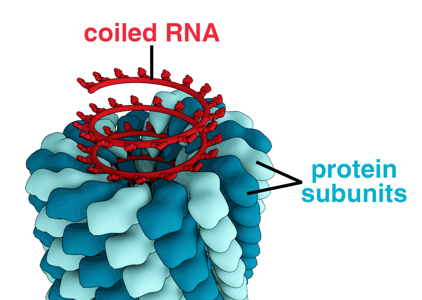
! It is the most throughly studied virus and was discovered by the Russain worker D. Ivanowsky (1892).
! It is rod shaped virus measuring 300 nm × 20 nm
! It is having helical symmetry.
! Having single stranded RNA which is 330 nm is length and having 7300 nucleotids.
! In a capsid number of capsomeres are 2130.
! 5% RNA and 95% protein ⇒ present in TMV.
Influenza virus :
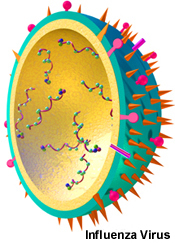
! Size 80 -120 nm , Spherical virus, infecting respiratory tract.
! Having helical symmetry, 10% RNA and 90% protein
! Having single stranded RNA, killed at 650C and active at low temperature.
! Crryptogram of influenza virus : R/1 : 2-3/10S/E : V/E
Bacteriophage Virus : Virus which infecting the bacteria
! Bacteriophage was discovered by F.W. Twort and Felix d’ Herelle
! Shell Singer explained that bacteriophage is made up of nucleoprotein (Nucleic Acid + protien)
! Hershy and Chase discovered heredity material - DNA in T2- bacteriophage through the radio tracer techniques.
Cyanophage : The virus which infects blue green algae are known as cyanophage. (Discovered by Safferman and Moris). Cyanophages contain ds DNA. The structure of cyanophages is similar to the bacteriophages. (ex Lpp-1 called so as it attacks Lyngbaya, Phormidium and Plectonema)
Sinsheimer : He discovered single stranded DNA in Ø × 174 bacteriophage.
IMP NOTE :
! In bacteriophages, generally DNA is present but in MS2 F2 r - 17 bacteriophages ss RNA is present.
! Generally DNA is double stranded but in Ø × 174 bacteriophage and in S13. E.coli phage, DNA is single stranded
Types of bacteriophages :
A. Prophages or non-virulent phages or non-infective phages : The phages which don’t cause lysis of bacteria are called prophages. Such bacterial cells which are having prophages inside them are called Lysogenic bacteria.
B. Virulent phages or infective phages or, Lytic phase : The phages which cause lysis of bacterial cell are once are called virulent phages.
IMP NOTE :
Most studied series of bacteriophages is T-series, i.e. T2, T4, T6 etc. (T-even phages are characterized by angular head and long contractile tail).
In T3 and T7 bacteriophage head is hexagonal.
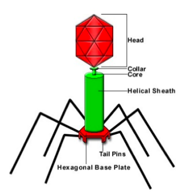
Structure of bacteriophages :

Having tadpole-like structure and differentiated into head & tail. Head is prism like having length 950 Å and breadth 650Å Tail is also 950 Å in length, joined to head by neck and collar. Tail is having hollow core of 80Å and is surrounded by tail sheath. At the end of tail, end plate is present to which 6 tail fibres are attached, each is 1500Å in length.
Function of tail fibres :
The tail fibres have two main functions :
(i) They help in the adsorption of phage particle on the surface of the bacterium
(ii) The enzymes secreted by these fibres are helpful in the lysis of bacterial cell wall.
IMP NOTE :
1. The water of Ganga is not spoiled due the presence of bacteriophage.
2. The circular area of dead bacteria on agar plate, is called plaque.
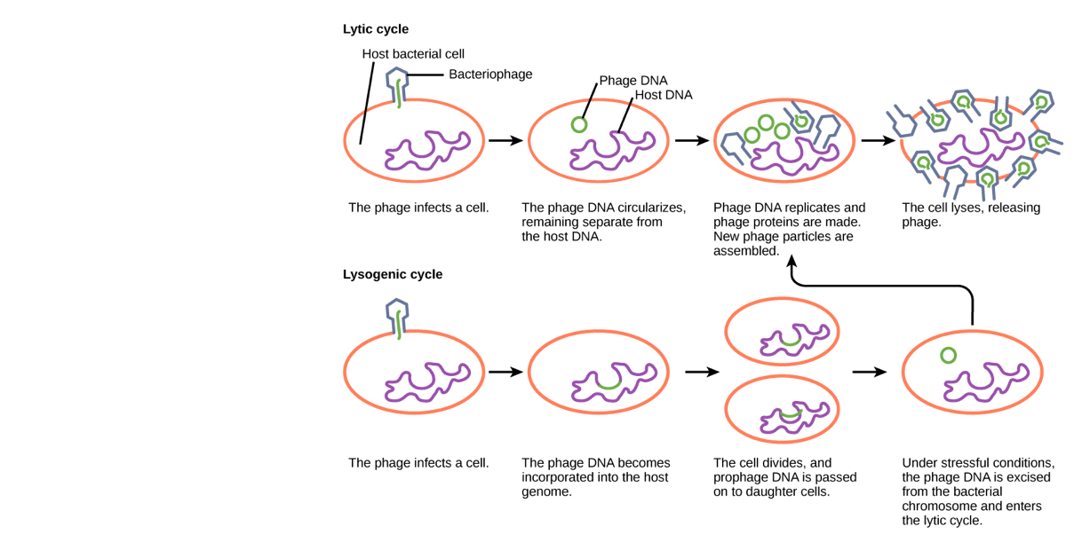
LIFE CYCLE OF BACTERIOPHASE :
The life cycle of bacteriophage is also known as infection cycle, which synthesizes many new phage particles thus, also referred as reproduction (or replication). The pages reproduce usually by two means - (i) Lysis (ii) Lysogeny
(i) Lysis / Lytic Ccle :
In this process virus gets like enzyme atached to the cell wall of bacteria at a specific place known as receptor site. At these receptor sites, Lysozyme synthesized by viruses react with bacterial cell wall. Consequently, a minute pore is formed through which DNA of the phage enters into the host cell. The empty capsid and tail fibres left behind are called ghost. After infection phage DNA assumes control of the cellular metabolism of bacterial cell and directs to synthesize the phage DNA and proteins. Subsequently, these new DNA molecules and protein particles assembled to form new bacteriophages which are liberated in the medium by the endolysis of host cell wall facilitated by the lysozyme like enzyme.
(ii) Lysogeny/Lysogenic cycle :
The initiator virus of this cycle is known as temperate phage/ \(\lambda\) - phage
The host cell is not degenerating in this cycle. The DNA of bacteriophage joined with the genophore of bacterium after the infection and replicates along with this. In this condition it is transmitted to progeny of bacteria. Such a virus called as provirus or Prophage. Bacteria which carry a provirus are called lysogenic bacteria and virus whose chromosomes becomes prophage are called lysogenic viruses.
If, it separates from the genophore artificially then it becomes virulent and start the Lytic cycle.
IMP NOTE :
1. It possible to induce lysogenic bacteria to lysis by irradiation with ultraviolet light or by exposure to some chemical like H2O2
2. Due to show reproductive process sometimes, millions of viruses can live their hosts for long period without any apparent indication of their presence. These are called latent or inapparent infections. [Multiplication of viral DNA takes place in the latent phage of virus]
Transduction :
When transfer of genetic material from one bacterium (Donor cell) to another bacterium (receptor cell) takes place by bacteriophage, called as transduction.
Discovered by - Zinder & Lederberg 1952) in Salmonella typhimurium.
Type of tansduction :
1. Generalised transduction : In this type of process bacteriophages are capable to transform any gene of bacteria.
2. Specialized transduction : In this type of process bacteriophages are capable to transform special part of donor genome.
Viroids
= T.O. Diener (1971) discovered some new infectious agents, which are still smaller than viruses. These subviral infectious agents are called viroids.
= Viroids contain only verylow mol. wt. RNA (ss RNA) and not protein coat.
= Viroids cause Potato spindle tuber disease (PSTV), Chrysanthemum stunt, Citrus exocortis, Cumumber pale fruit etc.
= Viroids cause peristent infectioins, i.e. never recovered/
IMP NOTE :
(i) Due to absence of protein coat viroids are also called as naked virus.
(ii) In virouds RNa, 246 to 388 nucleotides are present. They possesses the power of replication.
Virusoides :
Virusoides are like viroids, but are located inside the protein coat of a true virus, virusoid RNA can be either circular or linear. Virusoids are infectious by themselves because they are replicated only in the presence of their host.
Prions or Slow viruses :
! In 1966, three British scientists T. Alper, D. Haig and M. Clarke discovered infectious agents which are even smaller than viriod. They coined the term prion. But credit goes to professor Stanley B. Prusiner for the detailed study of Prions. Nobel prize was awarded to profesdor Prusiner in 1997 for his significant contribution.
! Prions lack their own genetic material (DNA or RNA). They are consisting of specific protein macro molecules which is known as prion protein or Prep.
! According to Prusiner, in most of the animals prion protein is generally associated with the chemical substance found in the nerve cells of barin.
! Prions are associated with Kuru (the laughing death) disease of man, Creutzfeldt jakob disease of humans and animals. Scrapie disease of sheep and goats, mad cow disease.
! Prion casue disease of mental disorder.
! In 1976 D.G. Gujdusek was awarded noble prize to the research of prion bases disease.
Interferons
! G.M. Findely and McCallum (1937) reported a phenomenon called viral interference in which the cell infected with one type virus becomes resistant to suerinfection by other viruses.
! Alliac Issacs and Lindeman (1957) gave the term interferons to the chemical substances responsible for viral interference.
! Interferons are produced by cells in mammals, rodents, birds, etc. and provide resistance against viruses.
! Hilleman and A. Tydall (1963) isolated interferon’s from hen’s egg infected with influenza virus.
! Interferons are protein molecules or polypeptides of low molecular weight which prevent Viral Multiplication.